Are you looking for a comprehensive guide on Pregnancy Ultrasound? You have landed on the right page. Getting an ultrasound test is one of the things that most thrills and terrifies you when you are pregnant. A Pregnancy Ultrasound is an essential part of any comprehensive prenatal care plan. Prenatal ultrasounds can reveal problems, track the mother’s and baby’s health, and evaluate the fetus’s development and growth.
During pregnancy, Pregnancy Ultrasound can be utilized for numerous purposes. If a previous ultrasound or blood test revealed an issue, your doctor may decide to request additional ultrasounds. The use of ultrasounds is discouraged by healthcare practitioners when there is no medical rationale or advantage, even though the technology is safe for both the mother and the kid. ER of Watuga is an industry-leading emergency room offering unmatched Pregnancy Ultrasound services in Texas and nearby areas. You can rely on us to acquire accurate ultrasound results.
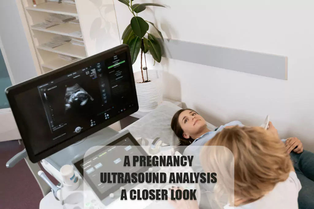
During a Pregnancy Ultrasound, your doctor or a qualified technician uses a plastic transducer to send high-frequency sound waves through your uterus. A computer receives these sound waves and uses them to create pictures of your unborn child. Keep reading to learn about the many kinds of Pregnancy Ultrasound, when they are most often ordered, and what to anticipate from your prenatal scans.
Pregnancy Ultrasound: What Is It?
A Pregnancy Ultrasound employs high-frequency sound waves to image both the developing fetus and the mother’s reproductive organs. The typical amount of Pregnancy Ultrasound performed changes from one pregnancy to the next.
Sonograms can detect abnormalities and track the progress of a healthy pregnancy. There are a variety of advanced ultrasounds available, including 3-D and 4-D models as well as fetal echocardiography, an ultrasound that examines the fetus’s heart in great detail.
Reasons for a Pregnancy Ultrasound
During pregnancy, Pregnancy Ultrasound can be utilized for numerous purposes. If your doctor found an issue in an earlier ultrasound or blood test, they may decide to perform additional ultrasounds. There are also non-medical uses for ultrasounds, such as showing the parents pictures or finding out the baby’s gender. The use of ultrasounds is discouraged by healthcare practitioners when there is no medical rationale or advantage, even though the technology is safe for both the mother and the kid.
The First Trimester of Pregnancy

In the first trimester of pregnancy (weeks one to 12), Pregnancy Ultrasound may be performed to:
- Confirm pregnancy
- Check the fetal heartbeat
- Determine the baby’s gestational age and predict its due date
- Check for multiple pregnancies
- Examine the placenta, uterus, ovaries, and cervix to determine ectopic pregnancy or miscarriage
- Check for any abnormal growth in the fetus
The Second and Third Trimesters of Pregnancy
Pregnancy Ultrasound may be performed in the second trimester (12 to 24 weeks) or the third trimester (24 to 40 weeks or delivery) to:
- Monitor the fetus’ development and position (breech, transverse, cephalic, or optimum)
- Determine the baby’s sex
- Confirm numerous pregnancies
- Examine the placenta for any issues, including placenta previa (when the placenta covers the cervix) and placental abruption (when the placenta separates from the uterus before delivery)
- Check for signs of Down syndrome (usually done between 13 and 14 weeks)
- Check for congenital anomalies or birth problems
- Examine the fetus for any structural abnormalities or blood flow issues
- Monitor the amounts of amniotic fluid
- Determine whether the fetus is receiving adequate oxygen
- Diagnose issues with the ovaries or uterus, such as pregnancy tumors
- Measure the cervix length to inform other procedures like amniocentesis and confirm intrauterine death
In a Pregnancy Ultrasound, What Can Be Detected?
The results of the Pregnancy Ultrasound scan will either be normal or not normal. If the test is normal, it indicates that your prenatal care provider did not detect any abnormalities and that your baby is developing normally. When your doctor notes an unusual result, it suggests something is wrong. In that case, your doctor would likely request further diagnostic testing or ultrasounds to rule out any potential issues.
If it is difficult to see all the structures required for that specific ultrasound, the Pregnancy Ultrasound may be incomplete. Occasionally, your healthcare practitioner may have trouble seeing what they need to do, due to your baby’s position or movement. In such instances, they will attempt again with a second Pregnancy Ultrasound. Some defects may not be detected until after birth because of the limitations of ultrasounds.
How to Prepare for An Ultrasound
A Pregnancy Ultrasound does not require any particular preparation. Some pregnancy care providers request that you arrive with a full bladder and not use the restroom before the Pregnancy Ultrasound. This improves the ultrasound image of your unborn child. This is a serious test that demands your undivided attention, so while a support person is welcome, we ask that you not bring children.
Although it isn’t typically necessary for abdominal ultrasounds, you could be requested to change into a hospital gown. In the first trimester of Pregnancy Ultrasound, if your doctor wants to do a transvaginal ultrasound, you’ll have to undress to the waist or wear a hospital gown.
What Happens During An Ultrasound
You will be lying on a comfortable table for the Pregnancy Ultrasound test. It’s easier for the person doing the ultrasound (or sonographer) to see the screen when the room isn’t very bright. A small amount of water-soluble liquid is put on the skin of your belly by the sonographer. It won’t hurt your skin or clothes, but the sticky substance might feel cold. Sound waves can travel farther with the help of this gel.
The sonographer will then put a sensor on your stomach skin. The sensor sends sound waves into your body, and your baby is one of the things that they bounce off. When sound waves bounce back, they make pictures on a screen. These pictures help your sonographer get important measurements, like the length and width of your baby’s head. They might draw lines on the screen or press a button to “freeze” certain views.
Having a prenatal scan doesn’t hurt very much. If you have to go to the toilet, you might feel a little pain. It takes about thirty minutes to finish the Pregnancy Ultrasound test. The only thing that is different about a transvaginal ultrasound is that the sensor is inside your vagina instead of on your belly.
Types of Pregnancy Ultrasounds
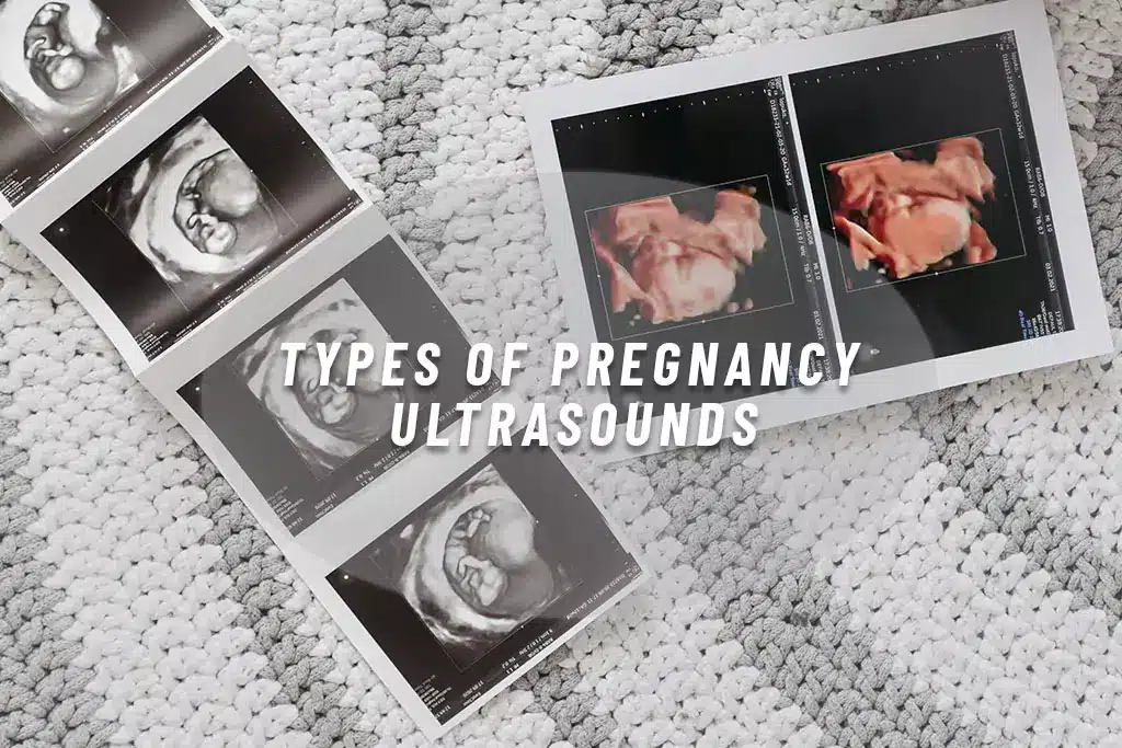
When a more thorough picture is needed, more advanced Pregnancy Ultrasound methods may be used. The doctor might be able to make a decision based on these if they find problems during your regular ultrasound.
3-D Ultrasound
3-D ultrasound is different from a 2-D ultrasound because it lets your doctor see the fetus and your organs’ width, height, and depth. This type of Pregnancy Ultrasound can help you figure out what’s wrong if you think something is wrong during your pregnancy. A 3-D ultrasound works the same way as a regular Pregnancy Ultrasound, but to make the 3-D picture, it needs a special probe and software. The mechanic also needs special training, so it might not be as easy to find.
4-D Ultrasound
A dynamic 3-D ultrasound is another name for a 4-D ultrasound. In contrast to other Pregnancy Ultrasounds, a 4-D ultrasound makes a video of the fetus that moves. It helps you see the baby’s face and move better. It also gets better blacks and highlights. This type of Pregnancy Ultrasound is done the same way as other ultrasounds but with different tools.
Transvaginal Ultrasound
To get a better picture, a transvaginal ultrasound may be used. This kind of Pregnancy Ultrasound is most likely to be used early in the pregnancy when it might be harder to get a helpful picture. A small ultrasound tool is put into the vagina for this test. While the pictures are being taken, the probe rests against the back of the genital area.
Fetal Echocardiography
If your doctor thinks your baby might have a congenital heart problem, a fetal echocardiography is done. This test might be done the same way as a regular pregnancy scan, but it might take longer to finish. Because it takes a picture of the heart of the fetus, it can show its size, shape, and structure. Your doctor can also see how your baby’s heart is beating through this form of Pregnancy Ultrasound, which can help them figure out if there are any heart issues.
How Many Ultrasounds Do You Have During Your Pregnancy?
During their pregnancies, the majority of women undergo one or two Pregnancy Ultrasounds. Your prenatal care physician and your specific health situation will determine the exact number and timing of these visits. Your healthcare practitioner may recommend more frequent Pregnancy Ultrasounds if they have reason to believe you or your baby is at high risk for complications during pregnancy.
How To Read Pregnancy Ultrasound Report?
Radiologists, cardiologists, and other professionals infer the images from the ultrasound scans performed by sonographers. Following these steps will make it easier for you to understand the Pregnancy Ultrasounds:
Locate your womb
Locating your womb should be your top priority. This is the first and most fundamental thing you need to know to read a Pregnancy Ultrasound. Looking at the thin white or light grey line that encircles the Pregnancy Ultrasound picture is usually all it takes to find the uterus. A big black patch, the amniotic fluid, may be seen inside these light grey and white lines. That your uterus may not encircle the entire ultrasound picture is something you should have in mind. Your doctor’s positioning of the probe during the ultrasound will determine this.
Take a Look at Your Baby

The second thing to observe during your Pregnancy Ultrasound is to locate your baby. The ultrasound picture shows the baby as white or grey. The darker area on the image represents the amniotic fluid, where the baby will be positioned. The ultrasound picture you see will show you your developing baby and the stage of your pregnancy. For instance –
- During the eighth week of pregnancy, when Pregnancy Ultrasound is typically performed, the size of the fetus is about equal to that of a baked bean
- The ultrasound pictures will reveal your baby’s head around the 12th week
- Moreover, if you look at the Pregnancy Ultrasound report after the 20th week of pregnancy, you will be surprised by the results. A baby’s vital organs, including its heart, feet, spine, eyes, and so on, are visible
How Much Does Ultrasound Cost?
The average cost of a Pregnancy Ultrasound may change based on the location in which state, or country the mother lives. The amount charged can also vary greatly depending on your provider. A doctor’s office or stand-alone clinic might not charge as much for a sonogram as a big hospital does because they have higher management costs. Call your source to find out how much your first ultrasound will cost. Stay in-network to keep your costs as low as possible.
Where Can I Get An Ultrasound Near Me?
Ultrasounds are frequent procedures, so you may easily find a professional near you. A doctor’s office, walk-in clinic, or hospital emergency room can all provide Pregnancy Ultrasound services. Because these are typically scheduled appointments, your doctor has the option of doing the operation in their office or recommending a nearby facility that is more convenient for you.
Ultrasounds can also be done in the emergency room. In such cases, they are usually employed to make a medical diagnosis or evaluate a change in a medical condition. But if you’re in so much pain or have noticed a dramatic shift in your body, you likely need to visit the emergency department. A preventative ultrasound may not be necessary if you practice proper hygiene.
ER of Watauga is a prominent healthcare provider offering urgent ultrasound services at reasonable prices in Fort Worth, Watauga, and nearby areas. We employ the best team of technicians and doctors having years of experience in the relevant field. You can rely on us to get unmatched Pregnancy Ultrasound test services. Get in touch with us today to book an appointment.
Conclusion
In conclusion, viewing a Pregnancy Ultrasound report might be nerve-racking for many expectant parents, but it is an important component of prenatal treatment. If you know about the different kinds of ultrasound scans, what to expect during the process, and how to read a Pregnancy Ultrasound report, you can feel less stressed and make sure that you and your baby get the best care possible. Talking to your doctor about any worries or questions you have is important because they can help you figure out what to do next in your pregnancy. ER of Watauga is your go-to emergency room for providing distinguished healthcare services in Texas and nearby areas. You can trust us to acquire the best Pregnancy Ultrasound at reasonable prices. Feel free to call us now or get a quote for a free consultation.
FAQs
What is the purpose of a Pregnancy Ultrasound?
During pregnancy, an ultrasound is used to check for problems, see how the baby is growing, find out how long the pregnancy has been going on, and get a general idea of the mother’s and baby’s health.
When is the best time to schedule a Pregnancy Ultrasound?
Between 18 and 20 weeks is the best time for an ultrasound, which is also called a “anatomy scan.” However, early ultrasounds in the first trimester may be done to make sure the pregnancy is viable and get an idea of when the baby will be born.
Are ultrasound procedures safe for the baby and the mother?
Yes, most people think that ultrasound treatments are safe. Sound waves, not radiation, are used in the technology, which has been studied in great detail. But it’s very important to follow the rules given and only get ultrasounds done by trained professionals.
What information can a Pregnancy Ultrasound provide?
An ultrasound during pregnancy can tell you a lot of useful things, like where the baby is, how fast it is growing, what organs are developing, and if any problems could happen. It helps doctors and nurses make smart choices about prenatal care and plans for delivery.
Can ultrasound determine the gender of the baby?
Yes, an ultrasound can often tell you what gender the baby is, usually between 18 and 20 weeks. It’s important to remember, though, that predicting the gender of a baby can be very inaccurate, and it’s not always possible because of things like the position of the fetus and the sonographer’s skill.