
Are you or one of your loved ones facing cardiac health issues and need to know everything about an ECG or EKG test? Welcome to the solution as we have got you covered through our detailed guide. An electrocardiogram is a fundamental medical diagnostic measure that traces the heart’s electrical activity on a time scale. ER of Watauga is your go-to emergency room for providing reliable diagnostic tests including ECG. We employ state-of-the-art tools and technology to provide unmatched findings for your diagnostic tests.
The heart’s various contractions are synchronized by natural electrical impulses to maintain proper blood flow. These impulses are captured by an ECG, which displays the heart rate, heart rhythm (steady or irregular), and the timing of the electrical impulses as they pass through the various cardiac chambers. Variations in an electrocardiogram may indicate several heart-related illnesses. It will be addressed in grave detail, about how a normal electrocardiogram works, how it should be read, and why it is a must for treating the common rhythms of heart diseases.
Understanding ECG
The abbreviation ECG, or electrocardiogram, stems from the original wording “Kardihographie” in German. It is a non-invasive diagnostic tool that is used to monitor heart rate, rhythm, and chamber size as well as damage, and the results of medications and pacemakers. In medical institutions like clinics and hospitals, this process is routine, a case in point being, the status of the cardiovascular system.
What Does a Normal ECG Look Like?
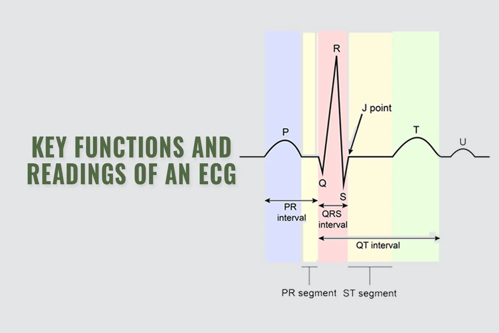
A normal ECG reading, which appears as a series of waves and lines, includes several distinct components reflecting different phases of the heart cycle:
- P Wave: Symbolizes atrial depolarization. Students are encouraged to share their opinions on different topics, outline solutions to real-world problems, and summarize the main points of a lecture.
- QRS Complex: Initiates the rate of quick acceleration and presents cardiac depolarization.
- T Wave: Represets the heart rate.
As for the specific variables of an electrocardiogram, their normal values vary a bit, but the accepted bounds, which are all-encompassing and agreed upon by the experts in the field, are the ones that best describe a healthy cardiac activity.
Key Functions and Readings of an ECG
ECG as it refers to a number of those pursuits provides deep knowledge of heart health
- Heart ailment diagnosis and monitoring: An electrocardiogram is an efficient tool to detect arrhythmia, coronary heart disease, the past of heart attack, and other diseases.
- When additional illnesses or ailments are present, check the health of your heart: for example, the cases of diabetes, genetically predisposed to heart disease, high blood pressure, high cholesterol, or smoking cigarettes.
- Electrocardiograms are important in this process as they can be used to track the heart’s circumstances when undergoing treatments and recovering from turned-up events like heart attacks by various cardiac treatments as well.
How to Read an ECG
Several steps are involved in reading an electrocardiogram:
- An electrocardiogram can return data on the milliseconds length of the cardiogram. That can vary from a minimum of 1 to 5 a minute depending on the type of heart rate.
- Assessing heart rhythm: Checking if there are consistent spaces between each complex wave/QRS (It is a complex formed by the R wave and the S wave).
- Observing the Cardiac Axis: In addition, we get to learn about the transmission pathway of the heart muscle plus much more.
- In this case, highlighting the QRS complex’s width, being the one which is larger than usual, and the P waves’ height, for instance.
- Recognizing Heart Attack Warning Signs: The classic example of this is ST segment depression found in a significant proportion of patients.
Advanced ECG Readings
ECG Leads and Positioning: The training of the best cardiac image is offered by the typical 12-lead electrocardiogram. To have a 3D model of the electrical signals coming from the heart, the electrocardiogram wires are placed in several distinguishable points on the patient’s chest and limbs.
How to Determine an ECG’s Heart Rate: Possibly, the electrocardiogram waveforms indicative of heart rate are calculated by multiplying the number of R waves observed within 6 seconds by a factor equal to 10.
The use of the term “borderline ECG” is ambiguous because it implies that the electrocardiogram test is neither special but also not clear cut. To establish a more stable diagnosis, further testing and checking might be required.
Conditions Detected by ECG
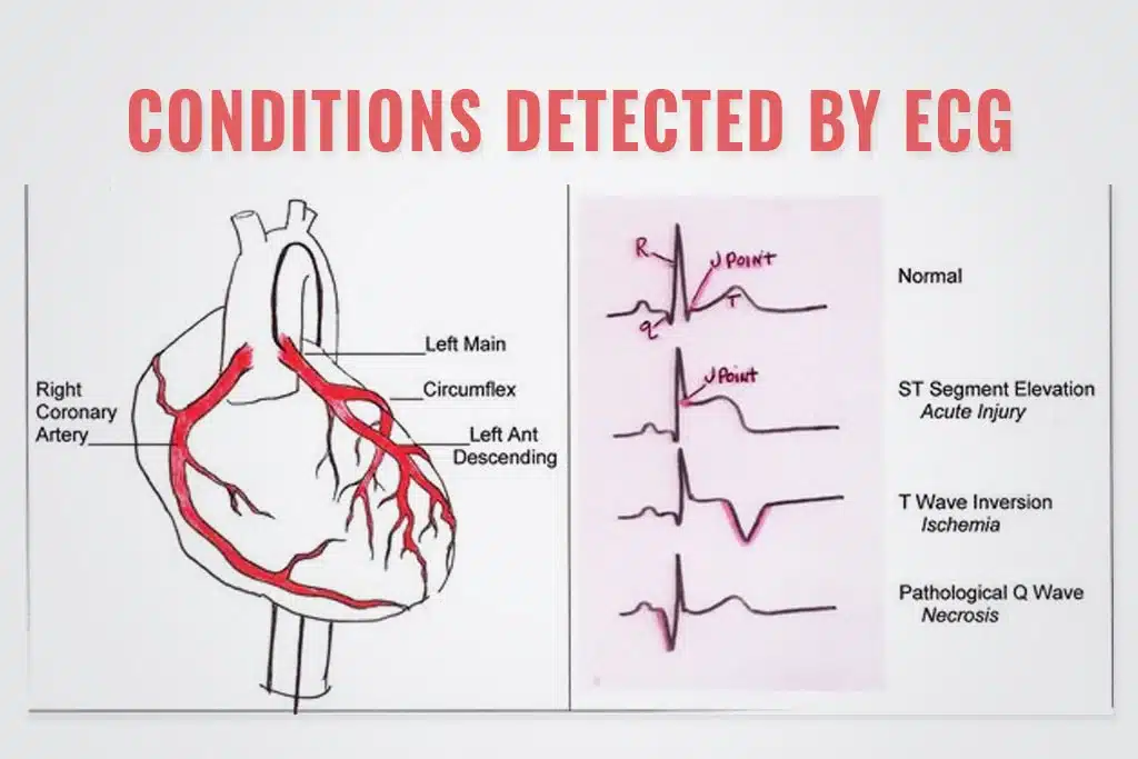
- Bradycardia Sinus ECG and Heart Disease ECG: Some subjects are being diagnosed with heart disease and have an established pattern in their electrocardiogram.
- Wolff-Parkinson-White, or WPW ECG: The changed electrocardiogram shows a wide, typical shortened phase interval of PR.
Dealing with Abnormal Results

- What causes abnormal ECG results: Examples may comprise pre-existing heart diseases, electrolyte disturbances, and post-heart-attack adjustments. Create your journey conference event in college.
- 5 lead ECG placement: It is also involved in telemetry (the functioning of cardiac rhythms), which could assist in identifying less frequently detected abnormal rhythms.
Conclusion
Mastering the reading and interpretation of an ECG or EKG may give crucial insights into an individual’s cardiac health, either now or in the future. Whether you are a patient looking to learn more about heart health tests or a medical professional improving your cardiology abilities, knowing how to interpret an electrocardiogram is crucial. At the ER of Watauga, we offer specialized care for patients facing cardiac health issues and ensure them their findings will be reliable and readable. You can trust us to get exceptional results for your diagnostic tests. We employ the best teams of physicians who can help you interpret the ECG test reports and suggest the best treatment plan ensuring the best cardiac health. Contact us now to make an appointment or to get further details.
FAQs
What is the borderline ECG?
An electrocardiogram with a borderline reading indicates that it is neither obviously abnormal nor normal. It may be necessary to conduct more monitoring or inquiry to rule out any underlying heart issues, but these modest anomalies may not suggest a severe health concern.
How is heart rate determined using an ECG?
The distance between two R peaks in a heart rate monitor pattern, or the R-R interval, can be used to calculate heart rate. Calculating 1500 divided by the number of little squares that fit between these R points is a simple method. Divide 300 by the total number of large squares that are located between the R points if you would prefer to use large squares. This indicates the heart rate in beats per minute.
In what location are the ECG lines placed for routine observation?
For a conventional 12-lead ECG to be measured correctly, the leads’ location is crucial. Precordial leads V1 through V6, or six of the leads, are positioned across the chest. The remaining six leads, referred to as limb leads, are situated on the arms and legs. The right leg does not record a pattern; it is a ground. The electrocardiogram will record all electrical activity from various locations around the heart provided it is positioned correctly. This is crucial for the diagnosis of several cardiac disorders.
What does a normal ECG look like?
An electrocardiogram, abbreviated ECG, demonstrates a consistent rhythmic pattern that provides information concerning the electrical activity of a heart in good health. Three distinct waves comprise the typical waveform: a T wave, a QRS complex, and a P wave. Atricular depolarization is denoted by the P wave, while ventricular depolarization is denoted by the QRS complex. Assigned to ventricular repolarization is the T wave. A typical electrocardiogram reading is distinguished by the inclusion of average intervals and amplitudes in these components; any noticeable deviation from these parameters cannot be detected.
What are some common causes of abnormal ECG results?
A wide array of medical conditions are capable of causing abnormal findings on electrocardiograms. The aforementioned conditions comprise arrhythmias (pericarditis included), ischemia (deficiency of blood supply to the heart), electrolyte imbalances, and heart attacks, among others. Electrocardiogram anomalies may also be caused by myocardial atrophy or other structural modifications to the heart, as well as complications related to preexisting cardiac conditions.